Instructions:
Normal Variants (cont.)
Another way or representing lead III (remember your algebra?) is:
III = aVF + (-aVL) Putting it visually:
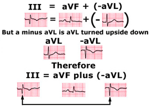
So an initial R wave in aVL which is quite common will translate into a Q wave in lead III. This Q wave can be very prominent at times but should not be misinterpreted as due to an inferior infarct. The same is true of the negative T wave in lead III. Notice in this case that a normal positive T wave in aVL is responsible for a negative T wave in lead III.
So, beware of lead III! For diagnosing an inferior infarct look first to lead aVF which DOES HAVE true spatial significance and after that to leads III and II to which aVF contributes, but only indirectly.
Case complete.